Ultra Sonography
Ultrasound can be enhanced by using Doppler technology which can measure whether an object is moving towards or away from the probe. This can allow the technician to measure blood flow in organs such as the heart or within specific blood vessels.
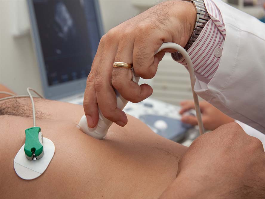
This technique helps to investigate clinical symptoms and gauge heart conditions, such as mumbles or impairment of the heart due to previous heart attack or infection. A heart ultrasound is a non-invasive way to detect the fitness of heart. Mostly this test carried out along an exercise stress test.
The image formed by the ultrasound machine is called a sonogram. Sonography is the procedure and ultrasound is the device. Sonography is organized by sonographers operating ultrasound devices. An echocardiogram is a graphic outline of the heart’s rhythm. During an echo test, ultrasound (high-frequency sound waves) from a hand-held stick positioned on chest, which provides pictures of the heart’s valves and chambers and assists the sonographer calculate the pumping performance of the heart.